TowardPi
YAlkaid
Description
Full Range Swept Source OCT, Next Generation
- Scan speed: 100.000 A-Scans / sec
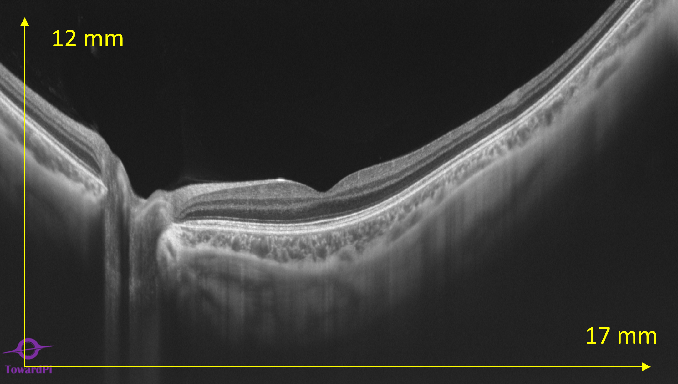
- Retina max scan (length X depth): 17 x 12 mm
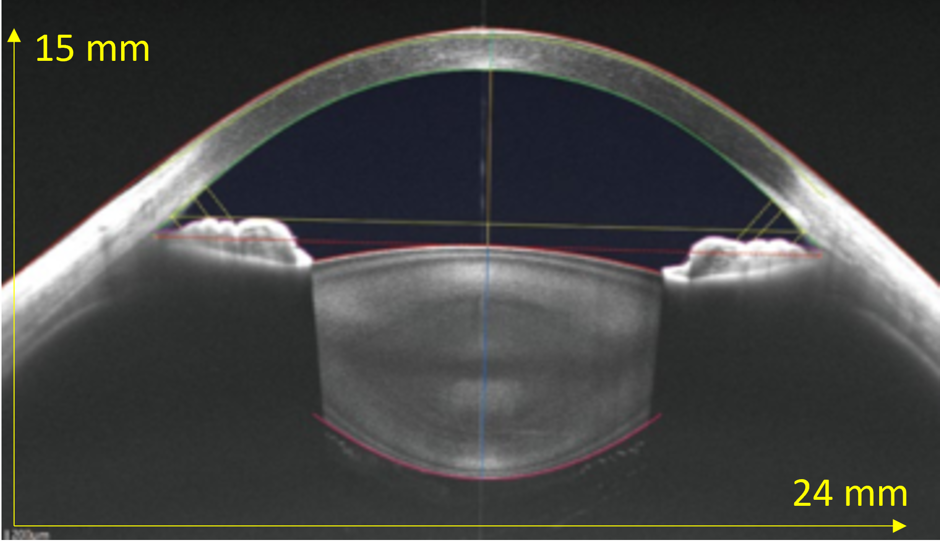
- Anterior segment max scan (length X depth): 24 x 15 mm
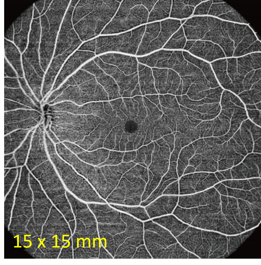
- OCT-A max. single scan size (Retina):15 X 15 mm
Full Range Retina OCT
- From posterior Vitreous to the Choroid-sclera
- 17 mm to 12 mm retina scan
- Automatic layer detection, segmentation and measurement of every layer of the retina and choroid
- Single line, Cross line, Grid Scan & Raster Scan
- LSO Fundus Image
Full range Anterior segment
- From Epithelium to the Posterior Capsule
- 24 mm to 15 mm scan
- Automatic identification of sclera spur and angle recess
- Anterior segment & AC Angle measurements
- Single line, HD Radial (8 lines) AS Radial (72 lines)
- Corna mapping: Pachymetry Map, Epithelium Map & Stroma Thickness Map (BL – Endothelium)
Full range OCT-A & En-Face OCT
- 3X3 mm, 6X6 mm, 12x12 mm, 15X15 mm in one shot and ultra wide field via montage
- Automatic segmentation in 7 layers: Vitreous, Superficial Vascular, Deep Vascular Complex, Retinal Vascular Layer, Avascular layer, Choroidal capillary layer, Choroidal vascular layer & Customizable segmentation
AI Based Choroid OCT-A with quantification tools
- Choroid: OCT-A, Vessel Index, Vessel Density, Vessel volume ratio (CVV/a), Vessel volume ratio (CSV/a), Choroidal Stroma Index
Comprehensive Glaucoma Analysis (with enhanced iHealth)
- 6x6mm Optical Nerve Head and Retinal Nerve Fiber Layer analysis resulting in cup area, rim area and cup/disc ratio
- 7x7mm Ganglion Macula Analysis measures the distance from internal limiting membrane (ILM) to outer inner plexiform layer IPL, which composes the inner 3 layers of the retina (NFL, ganglion cell layer (GCL) and inner plexiform layer), with significance map (GMA thickness vs. normative database).
- 6x6mm Disc with OCTA for blood flow analysis and density measurement of the blood vessels in the macula
- 15x9mm 3D scan for iHealth report for comprehensive analysis of the disc and macula
- Visualization of the pores of the lamina cribrosa

iSpot for PRP Laser management
- Unique solution to guide you in your photocoagulation treatment and evaluation
- Distinctive overlay of a retinal blood flow image with an en-face view highlighting laser treatment spots
LSO fundus imaging
- Line Scanning Ophthalmoscopy fundus imaging
3D OCT(-A) Retina, Optic Disc & AS
- Blood vessel visualization and quantification in all three zones
- Automatic Parameter measurement of thickness and angles in anterior segment
- Fundus Curvature measurement
Features:
- Scan speed: 100.000 A-Scans / sec
- Retina max scan (length X depth): 17 x 12 mm
- Anterior segment max scan (length X depth): 24 x 15 mm
- Fundus Imaging: LSO (Line-Scanning ophthalmoscope
- Min. pupil diameter: 2 mm
- Eye Tracking Speed: 60 Hz
- OCT-A max. single scan size (Retina):15 X 15 mm
- OCT-A max. single scan size (Anterior Segment): 18 x 18 mm
- OCT-A max. resolution (single scan): 1024 x 1024
- Anterior Segment quantification
- Thickness & Volume measurements: retina & choroid
- Glaucoma analysis: GMA, ONH, iHealth & Angle measurements
- Blood flow quantification: retina, choroid, optic disk and anterior segment
- Posterior curvature retina
- 3D structure & 3D vessels